A new database of potential antibiotic targets
Raleigh McElvery
January 20, 2021

Many cells, including bacteria, are covered in a sugar-rich coating that protects their membrane and internal components. These sugars are often joined to other macromolecules, like proteins or lipids, to form glycoconjugates. The glycoconjugates that encrust bacteria help prevent them from “popping” under environmental stress, and facilitate host-pathogen interactions. Because the sugary layer perpetuates survival and virulence, researchers are looking for ways to create chinks in this microbial armor — or better yet, to prevent it from being made in the first place.

Glycoconjugates are built by many enzymes working in close succession at the cell membrane. One enzyme family, comprised of phosphoglycosyl transferases (PGTs), is responsible for catalyzing the first step in the assembly line. Of this large enzyme family, one subtype in particular stands out: “monotopic” PGTs, which are unique to bacteria and could serve as antibiotic targets. If researchers can develop drugs that inhibit monoPGTs, the sugar armor wouldn’t be built and noxious bacteria could be easier to defeat.

new PNAS study co-authored by Professor of Biology and Chemistry, Barbara Imperiali, highlights the diversity and significance of these potential drug targets. Imperiali teamed up with graduate student Katherine O’Toole and Professor of Chemistry Karen Allen from Boston University to categorize over 38,000 different monoPGTs, compiling this information into the first database of its kind.

“We’ve taken an enzyme family that was once considered quirky and insignificant, and demonstrated that it’s actually very prevalent,” Imperiali says. “Hopefully the database will help us better understand these enzymes, their molecular pathways, and the human pathogens they support.”

Imperiali and her colleagues used sequence analysis of known monoPGTs to define a “signature” amino acid sequence. They leveraged this signature to identify the entire superfamily of monoPGTs amidst the 63,152 sequences downloaded from an online portal, which they then clustered into closely-related subtypes. The researchers also created a family tree, which included over 100 monoPGTs from diverse bacterial species. Imperiali hopes others will take advantage of this new information to pinpoint monoPGTs in pathogens of interest, and explore similarities and differences in related microbes and their enzymes.

The researchers’ analyses also revealed strange, new proteins that appeared to include two enzymes in one — a monoPGT fused to one of the other enzymes that typically play a separate role in the same sugar-modifying pathway. “It’s essentially one protein with two functions,” Imperiali explains. These fusion enzymes could reveal which enzymes “talk” to one another and work sequentially during the glycoconjugate-building process, she adds, revealing the complicated chain of events that creates the bacterial sugar shield.

The team even found cases where one monoPGT was fused to a member of a different PGT family — polytopic PGTs (polyPGTs). MonoPGTs and polyPGTs are involved in different pathways that each build glycoconjugates, so having a dual-function protein could allow cells to easily switch between mechanisms. Bacterial cells lack the organizational compartments that human and other eukaryotic cells have, so perhaps these fusion enzymes help exert control and order at different points in the cell cycle, Imperiali speculates. At the moment, though, the hybrid PGTs remain an evolutionary mystery.

While some researchers parse these ancient puzzles, others may use the database to inspire new drugs to combat antibiotic resistance. “At the end of the day,” Imperiali says, “we’ve shed light on a set of enzymes that could become pivotal therapeutic targets.”

Understanding antibodies to avoid pandemics

Structural biologist Pamela Björkman shared insights into pandemic viruses as part of the Department of Biology’s IAP seminar series.

Saima Sidik | Department of Biology
January 19, 2021

Last month, the world welcomed the rollout of vaccines that may finally curb the Covid-19 pandemic. Pamela Björkman, the David Baltimore Professor of Biology and Bioengineering at Caltech, wants to understand how antibodies like the ones elicited by these vaccines target the SARS-CoV-2 virus that causes Covid-19. She hopes this understanding will guide treatment strategies and help design vaccines against future pandemics. She shared her lab’s work during the MIT Department of Biology’s Independent Activities Period (IAP) seminar series, Immunity from Principles to Practice, on Jan. 12.

“Pamela is an amazing scientist, a strong advocate for women in science, and has a stellar history of studying the structural biology of virus-antibody interactions,” says Whitehead Institute for Biomedical Research Member Pulin Li, the Eugene Bell Career Development Professor of Tissue Engineering and one of the organizers of this year’s lecture series.

Immunology research often progresses from the lab bench to the clinic quickly, as was the case with Covid-19 vaccines, says Latham Family Career Development Professor of Biology and Whitehead Institute Member Sebastian Lourido, who organized the lecture series with Li. He and Li chose to focus this year’s seminar series on immunity because this field highlights the tie between basic molecular biology, which is a cornerstone of the Department of Biology, and practical applications.

“Pamela’s work is an excellent example of how fundamental discoveries can be intimately tied to real-world applications,” Lourido says.

Björkman’s lab has a long history of studying antibodies, which are protective proteins that the body generates in response to invading pathogens. Björkman focuses on neutralizing NetBet sportantibodies, which bind and jam up the molecular machines that let viruses reproduce in human cells. Last fall, the U.S. Food and Drug Administration (FDA) authorized a combination of two neutralizing antibodies, produced by the pharmaceutical company Regeneron, for emergency use in people with mild to moderate Covid-19. This remains one of the few treatments available for the disease.

Together with Michel Nussenzweig’s lab at The Rockefeller University, Börkman’s lab identified four categories of neutralizing antibodies that prevent a protein that decorates SARS-CoV-2’s surface, called the spike protein, from binding to a human protein called ACE2. Spike acts like the virus’s key, with ACE2 being the lock it has to open to enter human cells. Some of the antibodies that Björkman’s lab characterized bind to the tip of spike so that it can’t fit into ACE2, like sticking a wad of chewing gum on top of the virus’s key. Others block spike proteins from interacting with ACE2 by preventing them from altering their orientations. Understanding the variety of ways that neutralizing antibodies work will let scientists figure out how to combine them into maximally effective treatments.

Björkman isn’t satisfied with just designing treatments for this pandemic, however. “Coronavirus experts say this is going to keep happening,” she says. “We need to be prepared next time.”

To this end, Björkman’s lab has put pieces of spike-like proteins from multiple animal coronaviruses onto nanoparticles and injected them into mice. This made the mice generate antibodies against a mix of pathogens that are poised to jump into humans, suggesting that scientists could use this approach to create vaccines before pandemics occur. Importantly, the nanoparticles still work after they’re freeze-dried, meaning that companies could stockpile them, and that they could be shipped at room temperature.

Björkman’s talk was the second in the Immunity from Principles to Practice series, which was kicked off by Gabriel Victora from The Rockefeller University. Victora discussed how antibodies are produced in structures called germinal centers that are found in lymph nodes and the spleen.

Next in the series is Chris Garcia from Stanford University, who will speak on Jan. 19 about his lab’s work on engineering immune signaling molecules to maximize their potential to elicit therapeutic responses. To round out the series, Yasmine Belkaid from the National Institute of Allergy and Infectious Disease will speak on Jan. 26 about interactions between the gut microbiome and the pathogens we ingest. These talks complement a number of career development seminars that were organized by graduate students Fiona Aguilar, Alex Chan, Chris Giuliano, Alice Herneisen, Jimmy Ly, and Aditya Nair.

Biden taps Eric Lander and Maria Zuber for senior science posts

Lander to take a leave of absence to assume Cabinet-level post; Zuber to co-chair presidential advisory council.

Steve Bradt | MIT News Office
January 19, 2021

President-elect Joseph Biden has selected two MIT faculty leaders — Broad Institute Director Eric Lander and Vice President for Research Maria Zuber — for top science and technology posts in his administration.

Lander has been named Presidential Science Advisor, a position he will assume soon after Biden’s inauguration on Jan. 20. He has also been nominated as director of the Office of Science and Technology Policy (OSTP), a position that requires Senate confirmation.

Biden intends to elevate the Presidential Science Advisor, for the first time in history, to be a member of his Cabinet.

Zuber has been named co-chair of the President’s Council of Advisors on Science and Technology (PCAST), along with Caltech chemical engineer Frances Arnold, a 2018 winner of the Nobel Prize in chemistry. Zuber and Arnold will be the first women ever to co-chair PCAST.

Lander, Zuber, Arnold, and other appointees will join Biden in Wilmington, Delaware, on Saturday afternoon, where the president-elect will introduce his team of top advisors on science and technology, domains he has declared as crucial to America’s future. Biden has charged this team with recommending strategies and actions to ensure that the nation maximizes the benefits of science and technology for America’s welfare in the 21st century, including addressing health needs, climate change, national security, and economic prosperity.

“From Covid-19 to climate change, cybersecurity to U.S. competitiveness in innovation, the nation faces urgent challenges whose solutions depend on a broad and deep understanding of the frontiers of science and technology. In that context, it is enormously meaningful that science is being raised to a Cabinet-level position for the first time,” MIT President L. Rafael Reif says. “With his piercing intelligence and remarkable record as scientific pioneer, Eric Lander is a superb choice for this new role. And given her leadership of immensely complex NASA missions and her deep engagement with the leading edge of dozens of scientific domains as MIT’s vice president for research, it is difficult to imagine someone more qualified to co-chair PCAST than Maria Zuber. This is a banner day for science, and for the nation.”

Lander will take a leave of absence from MIT, where he is a professor of biology, and the Broad Institute, which he has led since its 2004 founding. The Broad Institute announced today that Todd Golub, currently its chief scientific officer as well as a faculty member at Harvard Medical School and an investigator at the Dana-Farber Cancer Institute, will succeed Lander as director.

Zuber, the E.A. Griswold Professor of Geophysics in MIT’s Department of Earth, Atmospheric, and Planetary Sciences, will continue to serve as the Institute’s vice president for research, a position she has held since 2013.

Separately, Biden announced earlier this week that he will nominate Gary Gensler, professor of the practice of global economics and management at the MIT Sloan School of Management, as chair of the Securities and Exchange Commission.

NetBet live casino

Eric S. Lander, 63, has served since 2004 as founding director of the Broad Institute of MIT and Harvard. A geneticist, molecular biologist, and mathematician, he was one of the principal leaders of the international Human Genome Project from 1990 to 2003, and is committed to attracting, teaching, and mentoring a new generation of scientists to fulfill the promise of genomic insights to benefit human health.

From 2009 to 2017, Lander informed federal policy on science and technology as co-chair of PCAST throughout the two terms of President Barack Obama.

“Our country once again stands at a consequential moment with respect to science and technology, and how we respond to the challenges and opportunities ahead will shape our future for the rest of this century,” Lander says. “President-elect Biden understands the central role of science and technology, and I am deeply honored to have been asked to serve.”

Trained as a mathematician, Lander earned a BA in mathematics from Princeton University in 1978. As a Rhodes Scholar from 1978 to 1981, he attended Oxford University, where he earned his doctorate in mathematics. Lander served on the Harvard Business School faculty from 1981 to 1990, teaching courses on managerial economics, decision analysis, and bargaining.

In 1983, his younger brother, Arthur, a developmental neurobiologist, suggested that, with his interest in coding theory, Lander might be interested in how biological systems, including the brain, encode and process information. Lander began to audit courses at Harvard and to moonlight in laboratories around Harvard and MIT, learning about molecular biology and genetics.

In 1986, he was appointed a Whitehead Fellow of the Whitehead Institute for Biomedical Research, where he started his own laboratory. In 1990, Lander was appointed as a tenured professor in MIT’s Department of netbet sports betting appBiology and as a member of the Whitehead Institute.

Lander’s honors and awards include the MacArthur Fellowship, the Breakthrough Prize in Life Sciences, the Albany Prize in Medicine and Biological Research, the Gairdner Foundation International Award of Canada, and MIT’s Killian Faculty Achievement Award. He was elected as a member of the U.S. National Academy of Sciences in 1997 and of the U.S. Institute of Medicine in 1999.

Maria Zuber

The daughter of a Pennsylvania state trooper and the granddaughter of coal miners, Maria T. Zuber, 62, has been a member of the MIT faculty since 1995 and MIT’s vice president for research since 2013. She has served since 2012 on the 24-member National Science Board (NSB), the governing body of the National Science Foundation, serving as NSB chair from 2016 to 2019.

Zuber’s own research bridges planetary geophysics and the technology of space-based laser and radio systems. She was the first woman to lead a NASA spacecraft mission, serving as principal investigator of the space agency’s Gravity Recovery and Interior Laboratory (GRAIL) mission, an effort launched in 2008 to map the moon’s gravitational field to answer fundamental questions about the moon’s evolution and internal composition. In all, Zuber has held leadership roles associated with scientific experiments or instrumentation on nine NASA missions since 1990.

As MIT’s vice president for research, Zuber is responsible for research administration and policy. She oversees more than a dozen interdisciplinary research centers, including the David H. Koch Institute for Integrative Cancer Research, the Plasma Science and Fusion Center, the Research Laboratory of Electronics, the Institute for Soldier Nanotechnologies, the MIT Energy Initiative (MITEI), and the Haystack Observatory. She is also responsible for MIT’s research integrity and compliance, and plays a central role in research relationships with the federal government.

“Many of the most pressing challenges facing the nation and the world will require breakthroughs in science and technology,” Zuber says. “An essential element of any solution must be rebuilding trust in science, and I’m thrilled to have the opportunity to work with President-elect Biden and his team to drive positive change.”

Zuber holds a BA in astronomy and geology from the University of Pennsylvania, awarded in 1980, and an ScM and PhD in geophysics from Brown University, awarded in 1983 and 1986, respectively. She has received awards and honors including MIT’s Killian Faculty Achievement Award; the American Geophysical Union’s Harry H. Hess Medal; and numerous NASA awards, including the Distinguished Public Service Medal and the Outstanding Public Leadership Medal. She was elected as a member of the National Academy of Sciences in 2004.

Todd Golub

Todd Golub, 57, will become the next director of the Broad Institute. He joined Dana-Farber and Harvard Medical School in 1997, and is currently a professor of pediatrics at Harvard Medical School and the Charles A. Dana Investigator in Human Cancer Genetics at Dana-Farber.

Golub served as a leader of the Whitehead Institute/MIT Center for Genome Research, the precursor to the Broad Institute. He has also been an investigator with the Howard Hughes Medical Institute, and has served as chair of numerous scientific advisory boards, including at St. Jude Children’s Research Hospital and the National Cancer Institute’s Board of Scientific Advisors.

Golub is also an entrepreneur, having co-founded several biotechnology companies to develop diagnostic and therapeutic products. He has created and applied genomic tools to understand the basis of disease, and to develop new approaches to drug discovery. He has made fundamental discoveries in the molecular basis of human cancer, and has helped develop new approaches to precision medicine.

“Broad is in a stronger scientific and cultural position today than at any point in our 16-year history,” Golub says. “Moreover, the pandemic has pushed us to think differently about nearly every aspect of how we collaborate and deliver on our scientific mission. We are well-positioned to work with the larger scientific community to confront some of the most urgent challenges in biomedicine: from developing novel diagnostics and therapeutics for infectious diseases and cancer, to understanding the genetic basis of cardiovascular disease and mental illness. I am honored to serve as director of this remarkable institution.”

Members of the Broad Institute’s Board of Directors thanked Lander for his lengthy service and expressed optimism in Golub’s ability to build upon that foundation.

“Todd’s deep knowledge of the Broad Institute community, its science, and its mission to propel the understanding and treatment of disease make him the perfect choice for the Institute’s next director,” says Louis Gerstner, Jr., chair of the Broad Institute Board of Directors. “Todd is well-positioned to lead the Institute and our key scientific collaborations forward, and the board is highly confident he will continue the Broad’s culture of innovation, collegiality, and constant renewal.”

Broad board member Shirley Tilghman, professor of molecular biology and public policy and president emerita of Princeton University, adds: “In its 16 years, the Broad has become one of the most unique institutions in the biomedical ecosystem. Under Eric’s and Todd’s leadership, it has developed powerful new methods and made many contributions to genomic medicine that will benefit human health.”

Why cancer cells waste so much energy

MIT study sheds light on the longstanding question of why cancer cells get their energy from fermentation.

Anne Trafton | MIT News Office
January 19, 2021

In the 1920s, German chemist Otto Warburg discovered that cancer cells don’t metabolize sugar the same way that healthy cells usually do. Since then, scientists have tried to figure out why cancer cells use this alternative pathway, which is much less efficient.

MIT biologists have now found a possible answer to this longstanding question. In a study appearing in Molecular Cell, they showed that this metabolic pathway, known as fermentation, helps cells to regenerate large quantities of a molecule called NAD+, which they need to synthesize DNA and other important molecules. Their findings also account for why other types of rapidly proliferating cells, such as immune cells, switch over to fermentation.

“This has really been a hundred-year-old paradox that many people have tried to explain in different ways,” says Matthew Vander Heiden, an associate professor of biology at MIT and associate director of MIT’s Koch Institute for Integrative Cancer Research. “What we found is that under certain circumstances, cells need to do more of these electron transfer reactions, which require NAD+, in order to make molecules such as DNA.”

Vander Heiden is the senior author of the new study, and the lead authors are former MIT graduate student and postdoc Alba Luengo PhD ’18 and graduate student Zhaoqi Li.

Inefficient metabolism

Fermentation is one way that cells can convert the energy found in sugar to ATP, a chemical that cells use to store energy for all of their needs. However, mammalian cells usually break down sugar using a process called aerobic respiration, which yields much more ATP. Cells typically switch over to fermentation only when they don’t have enough oxygen available to perform aerobic respiration.

Since Warburg’s discovery, scientists have put forth many theories for why cancer cells switch to the inefficient fermentation pathway. Warburg originally proposed that cancer cells’ mitochondria, where aerobic respiration occurs, might be damaged, but this turned out not to be the case. Other explanations have focused on the possible benefits of producing netbet sports bettingATP in a different way, but none of these theories have gained widespread support.

In this study, the MIT team decided to try to come up with a solution by asking what would happen if they suppressed cancer cells’ ability to perform fermentation. To do that, they treated the cells with a drug that forces them to divert a molecule called pyruvate from the fermentation pathway into the aerobic respiration pathway.

They saw, as others have previously shown, that blocking fermentation slows down cancer cells’ growth. Then, they tried to figure out how to restore the cells’ ability to proliferate, while still blocking fermentation. One approach they tried was to stimulate the cells to produce NAD+, a molecule that helps cells to dispose of the extra electrons that are stripped out when cells make molecules such as DNA and proteins.

When the researchers treated the cells with a drug that stimulates NAD+ production, they found that the cells started rapidly proliferating again, even though they still couldn’t perform fermentation. This led the researchers to theorize that when cells are growing rapidly, they need NAD+ more than they need ATP. During aerobic respiration, cells produce a great deal of ATP and some NAD+. If cells accumulate more ATP than they can use, respiration slows and production of NAD+ also slows.

“We hypothesized that when you make both NAD+ and ATP together, if you can’t get rid of ATP, it’s going to back up the whole system such that you also cannot make NAD+,” Li says.

Therefore, switching to a less efficient method of producing ATP, which allows the cells to generate more NAD+, actually helps them to grow faster. “If you step back and look at the pathways, what you realize is that fermentation allows you to generate NAD+ in an uncoupled way,” Luengo says.

Solving the paradox

The researchers tested this idea in other types of rapidly proliferating cells, including immune cells, and found that blocking fermentation but allowing alternative methods of NAD+ production enabled cells to continue rapidly dividing. They also observed the same phenomenon in nonmammalian cells such as yeast, which perform a different type of fermentation that produces ethanol.

“Not all proliferating cells have to do this,” Vander Heiden says. “It’s really only cells that are growing very fast. If cells are growing so fast that their demand to make stuff outstrips how much ATP they’re burning, that’s when they flip over into this type of metabolism. So, it solves, in my mind, many of the paradoxes that have existed.”

The findings suggest that drugs that force cancer cells to switch back to aerobic respiration instead of fermentation could offer a possible way to treat tumors. Drugs that inhibit NAD+ production could also have a beneficial effect, the researchers say.

The research was funded by the Ludwig Center for Molecular Oncology, the National Science Foundation, the National Institutes of Health, the Howard Hughes Medical Institute, the Medical Research Council, NHS Blood and Transplant, the Novo Nordisk Foundation, the Knut and Alice Wallenberg Foundation, Stand Up 2 Cancer, the Lustgarten Foundation, and the MIT Center for Precision Cancer Medicine.

Through my Viewfinder: savoring the little things
Irene Shih | MindHandHeart
January 13, 2021

If the coronavirus pandemic has taught me (and probably many of you) anything, it is that time is of the essence. Funnily enough, when campus and research were shut down, I had too much time that I didn’t know what to do with. In the beginning, I was glad that I could sleep in as much as I wanted but after a month of doing just that, I was so bored of staying indoors. One of the ways to get myself out of the house was the anticipation of witnessing something I would find endearing. I’ve always loved people watching and being observant of random details that most others miss. These little quiet acts, signs that life was happening inside these still, quiet facades, I say to them: I see you, I acknowledge your existence. I spent much of the spring and summer taking long walks around Cambridge, sometimes with a friend but mostly on my own. I feel happy that I managed to capture some of my favorite moments on these walks with my film camera.

My favorite part of photography is that I get to share how I see life, how I am able to note what matters to me. It has never been about creating a pretty image. What I find interesting, sad, beautiful, I take pictures of.


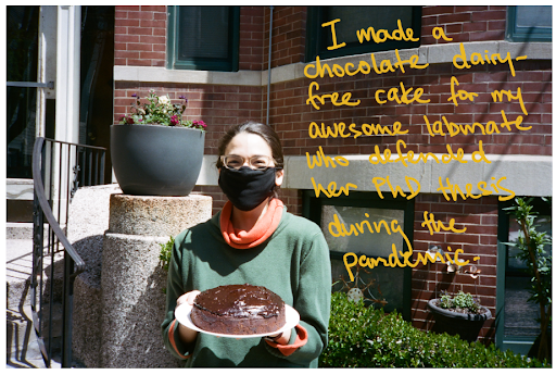
girl holding a cake

I made a chocolate dairy-free cake for my awesome labmate who defended her PhD thesis during the pandemic.

food cans on a table

In East Cambridge, free food laid out on a table outside a house.


wall with names
a blue house

I enjoyed walking around my neighborhood and seeing signs like these.

window in a house
window in a house
Neuroscientists identify brain circuit that encodes timing of events

Findings suggest this hippocampal circuit helps us to maintain our timeline of memories.

Anne Trafton | MIT News Office
January 12, 2021

When we experience a new event, our brain records a memory of not only what happened, but also the context, including the time and location of the event. A new study from MIT neuroscientists sheds light on how the timing of a memory is encoded in the hippocampus, and suggests that time and space are encoded separately.

In a study of mice, the researchers identified a hippocampal circuit that the animals used to store information about the timing of when they should turn left or right in a maze. When this circuit was blocked, the mice were unable to remember which way they were supposed to turn next. However, disrupting the circuit did not appear to impair their memory of where they were in space.

The findings add to a growing body of evidence suggesting that when we form new memories, different populations of neurons in the brain encode time and place information, the researchers say.

“There is an emerging view that ‘place cells’ and ‘time cells’ organize memories by mapping information onto the hippocampus. This spatial and temporal context serves as a scaffold that allows us to build our own personal timeline of memories,” says Chris MacDonald, a research scientist at MIT’s Picower Institute for Learning and Memory and the lead author of the study.

Susumu Tonegawa, the Picower Professor of Biology and Neuroscience at the RIKEN-MIT Laboratory of Neural Circuit Genetics at the Picower Institute, is the senior author of the study, which appears this week in the Proceedings of the National Academy of Sciences.

Time and place

About 50 years ago, neuroscientists discovered that the brain’s hippocampus contains neurons that encode memories of specific locations. These cells, known as place cells, store information that becomes part of the context of a particular memory.

The other critical piece of context for any given memory is the timing. In 2011, MacDonald and the late Howard Eichenbaum, a professor of psychological and brain sciences at Boston University, discovered cells that keep track of time, in a part of the hippocampus called CA1.

In that study, MacDonald, who was then a postdoc at Boston University, found that these cells showed specific timing-related firing patterns when mice were trained to associate two stimuli — an object and an odor — that were presented with a 10-second delay between them. When the delay was extended to 20 seconds, the cells reorganized their firing patterns to last 20 seconds instead of 10.

“It’s almost like they’re forming a new representation of a temporal context, much like a spatial context,” MacDonald says. “The emerging view seems to be that both place and time cells organize memory by mapping experience to a representation of context that is defined by time and space.”

In the new study, the researchers wanted to investigate which other parts of the brain might be feeding CA1 timing information. Some previous studies had suggested that a nearby part of the hippocampus called CA2 might be involved in keeping track of time. CA2 is a very small region of the hippocampus that has not been extensively studied, but it has been shown to have strong connections to CA1.

To study the links between CA2 and CA1, the researchers used an engineered mouse model in which they could use light to control the activity of neurons in the CA2 region. They trained netbet online sports bettingthe mice to run a figure-eight maze in which they would earn a reward if they alternated turning left and right each time they ran the maze. Between each trial, they ran on a treadmill for 10 seconds, and during this time, they had to remember which direction they had turned on the previous trial, so they could do the opposite on the upcoming trial.

When the researchers turned off CA2 activity while the mice were on the treadmill, they found that the mice performed very poorly at the task, suggesting that they could no longer remember which direction they had turned in the previous trial.

“When the animals are performing normally, there is a sequence of cells in CA1 that ticks off during this temporal coding phase,” MacDonald says. “When you inhibit the CA2, what you see is the temporal coding in CA1 becomes less precise and more smeared out in time. It becomes destabilized, and that seems to correlate with them also performing poorly on that task.”

Memory circuits

When the researchers used light to inhibit CA2 neurons while the mice were running the maze, they found little effect on the CA1 “place cells” that allow the mice to remember where they are. The findings suggest that spatial and timing information are encoded preferentially by different parts of the hippocampus, MacDonald says.

“One thing that’s exciting about this work is this idea that spatial and temporal information can operate in parallel and might merge or separate at different points in the circuit, depending on what you need to accomplish from a memory standpoint,” he says.

MacDonald is now planning additional studies of time perception, including how we perceive time under different circumstances, and how our perception of time influences our behavior. Another question he hopes to pursue is whether the brain has different mechanisms for keeping track of events that are separated by seconds and events that are separated by much longer periods of time.

“Somehow the information that we store in memory preserves the sequential order of events across very different timescales, and I’m very interested in how it is that we’re able to do that,” he says.

The research was funded by the RIKEN Center for Brain Science, the Howard Hughes Medical Institute, and the JPB Foundation.

Turning microbiome research into a force for health

A diverse group of researchers is working to turn new discoveries about the trillions of microbes in the body into treatments for a range of diseases.

Zach Winn | MIT News Office
January 8, 2021

The microbiome comprises trillions of microorganisms living on and inside each of us. Historically, some researchers have guessed at its role in human health, but in the last decade or so genetic sequencing techniques have illuminated this galaxy of microorganisms enough to study it in detail.

As researchers unravel the complex interplay between our bodies and microbiomes, they are beginning to appreciate the full scope of the field’s potential for treating disease and promoting health.

For instance, the growing list of conditions that correspond with changes in the microbes of our gut includes type 2 diabetes, inflammatory bowel disease, Alzheimer’s disease, and a variety of cancers.

“In almost every disease context that’s been investigated, we’ve found different types of microbial communities, divergent between healthy and sick patients,” says professor of biological engineering Eric Alm. “The promise [of these findings] is that some of those differences are going to be causal, and intervening to change the microbiome is going to help treat some of these diseases.”

Alm’s lab, in conjunction with collaborators at the Broad Institute of MIT and Harvard, did some of the early work characterizing the gut microbiome and showing its relationship to human health. Since then, microbiome research has exploded, pulling in researchers from far-flung fields and setting new discoveries in motion. Startups are now working to develop microbiome-based therapies, and nonprofit organizations have also sprouted up to ensure these basic scientific advances turn into treatments that benefit the maximum number of people.

“The first chapter in this field, and our history, has been validating this modality,” says Mark Smith PhD ’14, a co-founder of OpenBiome, which processes stool donations for hospitals to conduct stool transplants for patients battling gut infection. Smith is also currently CEO of the startup Finch Therapeutics, which is developing microbiome-based treatments. “Until now, it’s been about the promise of the microbiome. Now I feel like we’ve delivered on the first promise. The next step is figuring out how big this gets.”

An interdisciplinary foundation

MIT’s prominent role in microbiome research came, in part, through its leadership in a field that may at first seem unrelated. For decades, MIT has made important contributions to microbial ecology, led by work in the Parsons Laboratory in the Department of Civil and Environmental Engineering and by scientists including Institute Professor Penny Chisholm.

Ecologists who use complex statistical techniques to study the relationships between organisms in different ecosystems are well-equipped to study the behavior of different bacterial strains in the microbiome.

Not that ecologists — or anyone else — initially had much to study involving the human microbiome, which was essentially a black box to researchers well into the 2000s. But the Human Genome Project led to faster, cheaper ways to sequence genes at scale, and a group of researchers including Alm and visiting professor Martin Polz began using those techniques to decode the genomes of environmental bacteria around 2008.

Those techniques were first pointed at the bacteria in the gut microbiome as part of the Human Microbiome Project, which began in 2007 and involved research groups from MIT and the Broad Institute.

Alm first got pulled into microbiome research by the late biological engineering professor David Schauer as part of a research project with Boston Children’s Hospital. It didn’t take much to get up to speed: Alm says the number of papers explicitly referencing the microbiome at the time could be read in an afternoon.

The collaboration, which included Ramnik Xavier, a core institute member of the Broad Institute, led to the first large-scale genome sequencing of the gut microbiome to diagnose inflammatory bowel disease. The research was funded, in part, by the Neil and Anna Rasmussen Family Foundation.

The study offered a glimpse into the microbiome’s diagnostic potential. It also underscored the need to bring together researchers from diverse fields to dig deeper.

Taking an interdisciplinary approach is important because, after next-generation sequencing techniques are applied to the microbiome, a large amount of computational biology and statistical methods are still needed to interpret the resulting data — the microbiome, after all, contains more genes than the human genome. One catalyst for early microbiome collaboration was the Microbiology Graduate PhD Program, which recruited microbiology students to MIT and introduced them to research groups across the Institute.

As microbiology collaborations increased among researchers from different department and labs, Neil Rasmussen, a longtime member of the MIT Corporation and a member of the visiting committees for a number of departments, realized there was still one more component needed to turn microbiome research into a force for human health.

“Neil had the idea to find all the clinical researchers in the [Boston] area studying diseases associated with the microbiome and pair them up with people like [biological engineers, mathematicians, and ecologists] at MIT who might not know anything about inflammatory bowel disease or microbiomes but had the expertise necessary to solve big problems in the field,” Alm says.

In 2014, that insight led the Rasmussen Foundation to support the creation of the Center for Microbiome Informatics and Therapeutics (CMIT), one of the first university-based microbiome research centers in the country.

Tami Lieberman, the Hermann netbet online sports bettingL. F. von Helmholtz Career Development Professor at MIT, whose background is in ecology, says CMIT was a big reason she joined MIT’s faculty in 2018. Lieberman has developed new genomic approaches to study how bacteria mutate in healthy and sick individuals, with a particular focus on the skin microbiome.

Laura Kiessling, a chemist who has been recognized for contributions to our understanding of cell surface interactions, was also quick to joint CMIT. Kiessling, the Novartis Professor of Chemistry, has made discoveries relating to microbial mechanisms that influence immune function. Both Lieberman and Kiessling are also members of the Broad Institute.

Today, CMIT, co-directed by Alm and Xavier, facilitates collaborations between researchers and clinicians from hospitals around the country in addition to supporting research groups in the area. That work has led to hundreds of ongoing clinical trials that promise to further elucidate the microbiome’s connection to a broad range of diseases.

Fulfilling the promise of the microbiome

Researchers don’t yet know what specific strains of bacteria can improve the health of people with microbiome-associated diseases. But they do know that fecal matter transplants, which carry the full spectrum of gut bacteria from a healthy donor, can help patients suffering from certain diseases.

The nonprofit organization OpenBiome, founded by a group from MIT including Smith and Alm, launched in 2012 to help expand access to fecal matter transplants by screening donors for stool collection then processing, storing, and shipping samples to hospitals. Today OpenBiome works with more than 1,000 hospitals, and its success in the early days of the field shows that basic microbiome research, when paired with clinical trials like those happening at CMIT, can quickly lead to new treatments.

“You start with a disease, and if there’s a microbiome association, you can start a small trial to see if fecal transplants can help patients right away,” Alm explains. “If that becomes an effective treatment, while you’re rolling it out you can be doing the genomics to figure out how to make it better. So you can translate therapeutics into patients more quickly than when you’re developing small-molecule drugs.”

Another nonprofit project launched out of MIT, the Global Microbiome Conservancy, is collecting stool samples from people living nonindustrialized lifestyles around the world, whose guts have much different bacterial makeups and thus hold potential for advancing our understanding of host-microbiome interactions.

A number of private companies founded by MIT alumni are also trying to harness individual microbes to create new treatments, including, among others, Finch Therapeutics founded by Mark Smith; Concerto Biosciences, co-founded by Jared Kehe PhD ’20 and Bernardo Cervantes PhD ’20; BiomX, founded by Associate Professor Tim Lu; and Synlogic, founded by Lu and Jim Collins, the Termeer Professor of Medical Engineering and Science at MIT.

“There’s an opportunity to more precisely change a microbiome,” explains CMIT’s Lieberman. “But there’s a lot of basic science to do to figure out how to tweak the microbiome in a targeted way. Once we figure out how to do that, the therapeutic potential of the microbiome is quite limitless.”

Endowed funds to support MSRP-Bio
December 22, 2020

Dear colleagues,

I’m writing to share some really good news about our MIT’s Summer Research Program in Biology, or as most of us know it, MSRP-Bio. The simple take-home message: we now have endowed funds from Mike Gould and Sara Moss that will support about a dozen MSRP-Bio students each summer, for a very long time!

In 2015, Mike and Sara established the Bernard S. and Sophie G. Gould Fund to support students participating in MSRP-Bio. This gift was to provide opportunities to deserving students and to honor the memory of Mike’s parents.

Mike’s parents were both MIT alumni, and his father Bernie was a professor in the Biology Department from 1934 – 1987. Bernie and Sophie both committed their lives to supporting and counseling young students, and Mike and Sara chose to establish this fund to honor Mike’s parents and their deep and shared commitment to mentorship. Indeed, Mike and Sara share that commitment to support students and provide them with opportunities that could change their lives.

Mike and Sara have been remarkably dedicated to MSRP-Bio. Beginning with the first cohort of Gould Fellows in 2016, they visited each summer to meet and get to know these talented students. The first four meetings were in person, and then in the summer of 2020, the meeting was virtual due to the pandemic. Mike and Sara insisted on having the meeting, and even attended all of the student talks that summer. They have kept in touch with several of the Gould Fellow alumni and have gotten together with those who are in their hometown (NYC).

Mike and Sara have been so touched by the impact of their initial gift that they decided recently to provide additional support. To acknowledge this support and their commitment to our students and program, we are renaming MSRP-Bio the Bernard S. and Sophie G. Gould MIT Summer Research Program in Biology: BSG-MSRP-Bio, in honor of Mike’s parents.

We are deeply grateful to Mike and Sara for their commitment to and support for our community, their willingness to enable opportunities for students, irrespective of their specific research interests, and for the many talented individuals who will benefit from the experiences afforded by their generous gift. 

This gift is a great way to end the year. Wishing everyone a wonderful holiday season and a Happy Healthy New Year!

Best wishes,
Alan Grossman, Department Head

Ruth Lehmann receives 2020 Francis Amory Prize
Greta Friar | Whitehead Institute
December 21, 2020

Whitehead Institute Director Ruth Lehmann has been awarded the 2020 Francis Amory Prize in Reproductive Medicine and Reproductive Physiology by the American Academy of Arts & Sciences. Lehmann shares the prize with geneticist Gertrud M. Schüpbach, emeritus professor at Princeton University and a longtime friend and colleague. The Amory Prize recognizes outstanding achievements in medicine and reproductive physiology, and Lehmann and Schüpbach are being recognized for their contributions to areas including DNA repair, embryonic development, RNA regulation, and stem cell research. Lehmann, who is also a professor at the Massachusetts Institute of Technology, studies the biological origins of germ cells, the sex cells that produce eggs and sperm. Her research has shed light on many aspects of the germ cell life cycle, including how germ cells first form and become set apart from the rest of the body’s cells, how they migrate to the gonads during embryonic development, and how they remain protected in order to produce the next generation, preserving and passing on the complex instructions to construct a new life. Lehmann and Schüpbach will accept the Amory Prize at a virtual American Academy of Arts & Sciences event on February 3, 2021. To learn more, click here.

Disarming cancer
Greta Friar | Whitehead Institute
December 21, 2020

Cancer is at its most deadly when two things occur: the cancer cells metastasize, spreading to new sites in the body, and the cells become resistant to treatment. The epithelial-mesenchymal transition (EMT) is a process that cancer cells may undergo that enables them to do both of these things. Cells that undergo this process are called “quasi-mesenchymal” cancer cells, and they are mobile, aggressive, and harder to kill. They can resist attacks launched both by the body’s own immune system as well as immune checkpoint blockade therapy (ICB), an increasingly employed clinical treatment that works by liberating cells of the immune system from certain constraints, thereby allowing them to attack cancer cells. Anushka Dongre, a postdoctoral researcher in the lab of Whitehead Institute Founding Member Robert Weinberg, had previously found that even a small population of quasi-mesenchymal NetBet sportcells within a mouse breast cancer tumor—as little as 10% amongst a majority of cells that had not gone through the EMT—could protect the entire tumor from a version of ICB called anti-CTLA4 therapy. Most breast cancers in humans contain some minority populations of quasi-mesenchymal cells, as do many other types of human tumors, likely contributing to ICB therapy’s mixed success rates in the clinic.

Because cells that have been through the EMT process play such a large role in making cancers more deadly and less responsive to treatment, Dongre set out to understand how to defang them. Her first step was to figure out how minority populations of quasi-mesenchymal cells within a breast tumor make the tumors as a whole resistant to immune therapy. Then she studied how to disable those mechanisms. The work, described in a paper published in Cancer Discovery on December 16, includes studies in mice showing that disabling those resistance mechanisms can sensitize otherwise-resistant tumors to anti-CTLA4 checkpoint blockade immunotherapy and reduce the severity of metastasis.

Dongre had previously studied how quasi-mesenchymal cells alter the area in and around a tumor to render it more favorable for the outgrowth of a cancer. They keep out of the core of the tumor the type of immune cells that can destroy cancers, and instead let in other types of immune cells that the tumor is able to co-opt to its benefit, thereby protecting it from immune attack. 

In her latest research, Dongre identified six molecules that quasi-mesenchymal cells produce and release that help them perturb the tumor’s surroundings, protecting cells throughout the tumor from immune attack and elimination. She then tested what happened when the release of each of the protective molecules was suppressed. She discovered that eliminating release of either of two molecules, CSF1 and SPP1, made the tumors significantly more susceptible to the immune attack and thus elimination by ICB therapyHoweverthe strongest therapeutic benefit came when she prevented production of CD73, an enzyme usually made by the quasi-mesenchymal cells that produces the immunosuppressive molecule adenosine. In mice, anti-CTLA4 therapy was very effective against tumors in which CD73 and thus adenosine had been eliminated from the quasi-mesenchymal cells, in some cases, succeeding in eliminating the tumors entirely. These findings are consistent with previous research that identified CD73 as a good complementary target for immunotherapy. Furthermore, the experiments demonstrated the utility of combining anti-CD73 therapy with anti-CTLA4 immunotherapy in order to successfully treat tumors that would usually not respond to treatment by ICB therapy alone. Dongre was particularly excited to see the combination of anti-CD73 and anti-CTLA4 reduce the number and size of metastatic tumors.

Dongre hopes that these insights will prove useful for patients.

“There is this minority population of mesenchymal cells present in many patient tumors, creating a big barrier to therapy. I’m hopeful that by identifying the drivers that can sensitize this population to treatment, our work can one day help patients suffering from cancers that are resistant to current therapies,” Dongre says.